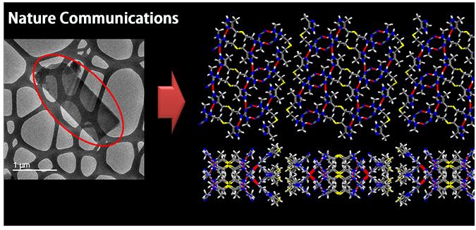
2019年8月6日, 日本电子株式会社的NMR专家西山博士(YUSUKE NISHIYAMA)在Nature Communications上发表了一篇论文:Understanding hydrogen-bonding structures of molecular crystals via electron and NMR nanocrystallography。文章中阐述使用TEM和固体NMR可以成功地对100nm-1um的纳微晶体包括氢键结构进行表征, 文章中注明所使用的设备为: JEOL 的透射电子显微镜(型号 JEM-2200FS)和JEOL的固体核磁共振(型号 JNM-ECZ600R)。
具体详情请登陆下面网址:
https://www.nature.com/articles/s41467-019-11469-2
摘要请登录:http://www.jeol.com.cn/app/index
Visualization of hydrogen-bonding: Electron and NMR nano-crystallography
Product used :Transmission Electron Microscope (TEM), nuclear magnetic resonance (NMR)
Crystalline structure including hydrogen atoms are now available from nano-to micro-crystals of 100 nm to 1 μm using electron and NMR nano-crystallography approach. The overall crystalline structures can be determined by electron diffraction (ED) which is one of the observation mode of transmission electron microscope (TEM). However, the ED based structures have several problems including 1) invisible hydrogen atoms and 2) mis-assignment of carbon, nitrogen and oxygen atoms. The former brings crucial problems to understand hydrogen bonding networks and the latter results in ambiguous molecular conformation. On the other hand, solid-state NMR can directly observe 1) hydrogen (1H) and 2) carbon (13C) and nitrogen (14N, 15N). Here, we combine ED and solid-state NMR through the first principle quantum computation with the NMR crystallography approach for crystalline structure solution. The method, electron and NMR nano-crystallography, can be applied to nano-to micro-crystals even for mixture samples. First, we have demonstrated the structure solution of L-histidine, whose structure is already known, as a proof of concept. Then, we have solved the crystalline structure of cimetidine form B, whose structure was previously unknown.
The electron and NMR nano-crystallography can be applied to many pharmaceutical samples including pharmaceutical formulation as well as systems, in which only nano-to micro-sized crystals are available, including PCP/MOFs.

Figure: TEM image of cimetidine form B and crystalline structure solved by electron and NMR nano-crystallography.The cimetidine form B is needle shape crystals (indicated by red circle on the left figure) and often fails to form a large single crystal for single crystal X-ray diffraction. In addition, the contamination of form C is often observed, hampering the structural solution by powder X-ray diffraction. Here, we solved the crystalline structure of cimetidine form B for the first time.
C. Guzmán-Afonso†, Y.-l. Hong†, H. Colaux, H. Iijima, A. Saitow, T. Fukumura, Y. Aoyama, S. Motoki, T. Oikawa, T. Yamazaki, K. Yonekura& Y. Nishiyama*, Nat. Comm. in press DOI: 10.1038/s41467-019-11469-2


